Super Resolution Microscope: Lattice SIM/STED/STORM
Description
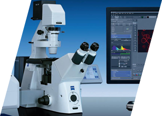
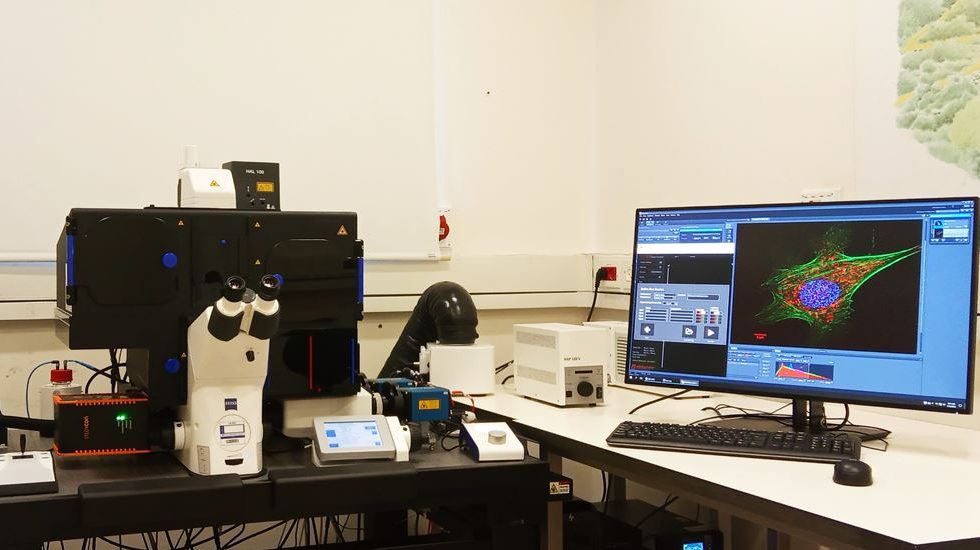
Multi Modality Super Resolution (SR) microscope that combines lattice SIM, 2D-STED and STOTM/PALM methods for SR, this system is designed for live-cell imaging.
The Elyra 7 eLS is a flexible platform featuring unique lattice structured illumination (SIM^2) for fast and gentle 3D super-resolution on live samples as well as Single Molecule Super-resolution imaging. Lattice SIM^2 provides optical sectioning and a doubling of diffraction-limited resolution in 3D (60 nm in xy and 150 nm in z). SMLM part allows for resolutions in the typical range of 20-30 nm laterally and 50-80 nm axially. The system features groundbreaking light efficiency and allows for gentle live cell imaging at speeds of dozens of frames per second over time. It includes state-of-the-art sCMOS cameras for large field of view imaging, SIM- and SMLM optimized optics, focus drift compensation control and full incubation.
The STEDYCON is a laser scanning confocal microscope and STED nanoscope with a resolution down to 30 nm. It has a super-intuitive user interface.
Features
SYSTEM SPECIFICATIONS:
- Super Resolution (SR) imaging: Lattice SIM^2 (Zeiss), STORM (Zeiss) and STED (Abberior)
- Confocal imaging : StedyCon (Abberior- confocal)
- Z Stack imaging: Apotom (Zeiss)
TIRF imaging:
- Stand: Zeiss Axio Observer 7 SR RP stand for ELYRA 7
- XY-stage: motorized
- Z – Z PIEZO WSB 500 um range
- Definite Focus 2 (Zeiss)
- StedyFocus (Abberior)
- HXP fluorescence illumination
- Equipped for live imaging (temperature and CO2 incubation)
- Software: ZEN
LASER OPTIONS
The Zeiss system includes 4 visible solid state lasers:
- UV diode laser – 405 nm (50mW)
- Blue diode laser 488 (500mW)
- Green diode laser – 561 nm (500mW)
- Red diode laser – 642 nm (500mW)
The Abberrior system includes 4 visible solid state lasers:
- UV diode laser – 405 nm (continues)
- Blue diode laser 488 (pulse)
- Green diode laser – 561 nm (pulse)
- Red diode laser – 640 nm (pulse)
- STED laser depletion: 775nm (pulsed)
CAMERA SPECIFICATIONS
- Model: Two pco.edge sCMOS (version 4.2 CL HS)
- Effective number of pixels: 2048 x 2048
- Pixel size: 6.5µm x 6.5µm
- Effective Area: 13.312mm x 13.312mm
- Exposure times from 100 µs to 20 s
- Dynamic range: 16 bit
- Max frame rate: 100 fps @ full frame (CL/CLHS)
- 40 fps @ full frame (USB)
- Quantum efficiency (peak): > 82% (@590nm)
- Read out noise: extreme low readout noise of 0.8 e.
OBJECTIVES
| Magnitude | Objective | Serial # | NA | Immersion | Working distance (um) | Imaging Application |
|---|---|---|---|---|---|---|
| 10X | EC Plan-Neofluar | 420340-9901-000 | 0.3 | DRY | 5200 | |
| 40X | Plan-Apochromat | 420762-9900-000 | 1.4 | Oil | 130 | Apotome |
| 63X | Plan-Apochromat | 421787-9970-799 | 1.2 | Water | 280 | Lattice SIM |
| 63X | Plan-Apochromat | 420782-9900-799 | 1.4 | Oil | 190 | Lattice SIM |
| 63X | Alpha Plan-Apochromat | 420780-9971 | 1.46 | Oil | 100 | Lattice SIM/ STED |
| 100X | Alpha Plan-Apochromat | 420792-9800-720 | 1.46 | Oil | 110 | STORM/PALM/ STED/TIRF |
CAMERA FILTERS
| Beam Splitter | LP 560 | BP490-560/ LP640 | ||
|---|---|---|---|---|
| CAMERA | CAM1 | CAM2 | CAM1 | CAM2 |
| Detection | BP 570-620 | BP 420-480 | BP 495-550 | BP 420-480 |
| Detection | LP 655 | BP 495-550 | LP 655 | BP 570-630 |
| Detection | — | — | — | LP 740 |
Acquisition & Analysis
- Acquisition is performed using the ZEN black software which provides wide range of image processing functions: 2D/3D, projection, reconstructing, co localization, intensity measurements and more. The flexible secondary dichroic beam splitter makes this system fully capable of acquiring super resolution and performing dual camera imaging. In addition, Elyra 7 with Lattice SIM^2 takes you beyond the diffraction limit of conventional microscopy to image your samples with super-resolution. Elyra 7 is the fastest processes in living samples – in large fields of view, in 3D, over long time periods, and with multiple colors. Imaging with incredibly high speed – at 255 fps. Elyra 7 combine Lattice SIM^2 with single molecule localization microscopy (SMLM) for techniques such as PALM, dSTORM and PAINT.
- The STEDYCON software has a user friendly GUI that required only several minutes of training to acquire STED and confocal images.
Applications
The system enables amongst many others, the following applications:
- Super-Resolution Imaging:
Lattice Structure Illumination Microscopy (Lattice SIM^2)
STimulated Emission Depletion (STED) Microscopy :2D STED
Single Molecule Localization Microscopy (SMLM): PALM/dSTORM/PAINT - Section Imaging (Z stack):
Apotome -Zeiss
Confocal -Abberior - TIRF- Total Internal Reflection Fluorescence Microscopy
- Fast Image Acquisition for Live Cell Imaging and Time-Lapse studies
- Z-Stacks for localization of cells in living tissues and 3-D reconstruction
- Super-resolution localization of sub-cellular compartments and quantities co-localization
- FRET – fluorescence resonance energy transfer
- FRAP – fluorescence recovery after photobleacing in a super-resolution microscopy
- PhotoActivation and PhotoManipulation in a super-resolution microscopy
* Mark and find feature – The motorized stage allows marking specific coordinates using small magnifications or tiling of large scanning areas for further detailed scanning.


